
| |
There are 3 families of probes which can be used with the confocal
microscope in the AIF. They are:
Green (e.g. eGFP, FITC, Cy2, Calcium Green, Alexa 488)
Red (e.g. Rhodamine, Texas Red, Cy3, Alexa 568, Propidium Iodide)
Near Infra-red (e.g. Cy5, Syto 60, Toto-3)
[Read more about probes here]
STAINING WITH Cy5, propidium iodide and FITC
from the laboratory of Dr. Phyllis Novikoff
Department of Pathology, AECOM
(718) 430-2028 novikoff@aecom.yu.edu
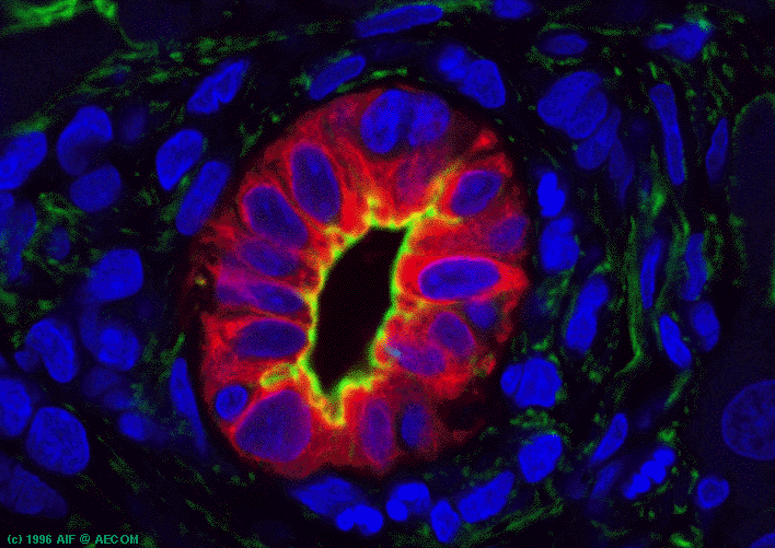
| This single optical section collected with the confocal microscope from a
thick vibratome section is from a slice of rat liver with a crossection through a bile
ductule. Nuclei were stained with propidium iodide and are represented here in blue. The large nucleus in the lower right (cut off by the edge of the image) belongs to a hepatocyte. F-actin was stained with FITC phalloidin and is represented here in green. The apical surface of the bile ductule cells is actin rich. Also, a bile canaliculus is visible in the far upper left corner. An intermediate filament specific to the bile ductule cells in this tissue is stained by immunocytochemistry with Cy5 and is represented here in red. Image was collected with a BioRad MRC 600 LSCM which was in the AIF from 1991 through 2000. The MRC 600 has been upgraded to a Radiance 2000. |
For more on this image, please refer to Figure 3 in
Novikoff, P.M., Yam, A. and Oikawa, I. (1996) Blast-like cell compartment in
carcinogen-induced proliferating bile ductules. American Journal of Pathology.
148(5):1473-1492.
![]()
![]()