| |
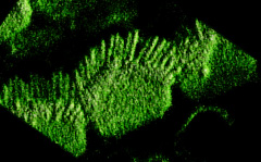
Inner ear hair cell imaged by confocal microscopy. Single optical sections collected with the BioRad MRC 600 of cells stained for f-actin were reconstructed using VoxelView to produce these volume renderings with lighting convolutions to appear similar to scanning electron micrographs. Cells courtesy of Dr. H. Staecker.