| |
| CRYOmicroscopy |
Cryo transmission electron microscopy (cryoTEM) is used with vitreous ice-embedded samples as a quantitative tool to investigate the assembly, regulation and function of biologically important macromolecular complexes. Despite continuous progress in x-ray crystallographic and NMR methods, it is still difficult using these techniques alone to obtain atomic level structural information about the many complex macromolecular assemblies that govern fundamental cell biological processes. It is now clear that a powerful approach is to combine structural information obtained at different levels. The structural information obtained from cryoTEM and computer based 3-D reconstruction spans the gap between our ability to obtain structural information about large macromolecular assemblies using conventional light and electron microscopy, and high-resolution data obtained from NMR or X-ray crystallography. These data can be combined to build up a detailed picture of the overall architecture of the complexes and the interactions between the components.
The development of an electron cryomicroscopy laboratory is a major component of our ongoing low temperature electron microscopy initiative. The focus of the electron cryomicroscopy laboratory will be analysis of macromolecules frozen in vitreous ice. Another application of the newly installed 200KV electron cryomicroscope will be electron tomography. We will be able to construct three-dimensional models of ultrastructural morphology.
The establishment of this electron cryomicroscopy laboratory is another step in our continuing effort to obtain better morphological preservation with less chemical processing. The AIF has a long history of low temperature techniques for electron microscopy, beginning with freeze fracture in the late 'seventies. Five years ago Frank Macaluso introduced metal mirror quick-freeze fixation which could be followed by freeze substitution or high resolution rotary shadowing. Three years ago we began to develop expertise in cryo ultra thin sectioning with the goal of preserving fine structure while maintaining epitope conformation for immunogold labeling. The point of all this effort is that chemical fixation proceeds slowly compared with quick freezing methods that immobilize cells in milliseconds. The addition of electron dense stains and metal shadowing further obscure fine structure as they enhance contrast under the electron beam. We now have the ability to directly view frozen hydrated samples in the electron microscope.
To further development of low temperature methods at AECOM, Dave Hall and Frank, along with a Major User Group that included Dave Sharp, Peter Mundel, Rob Singer, John Condeelis, Dick Hays and Michael Lisanti, applied for a Shared Instrumentation Grant in March 2002 to purchase a high-pressure freezer. The advantage of HPF over other quick-freeze techniques is the ability to preserve to a depth of more than 200 microns in tissue without ice crystal damage. In addition, the HPF will enable us to preserve samples that have been traditionally difficult to fix chemically such as C. elegans, drosophila embryos and yeast. [The HPF grant was approved and funded; the AIF now offers HPF as a service. -Ed. Mar. 2005]
The wide range of low temperature techniques makes the Analytical Imaging Facility a unique laboratory in New York City.
The staff of the AIF looks forward to facing the challenges in developing this technology so that our facility excels in low temperature electron microscopy of all types. We look forward to helping you apply the technology in your research.
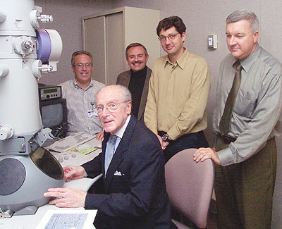
Opening Reception, October 2002
Introduction by Dr. John Condeelis: Today we meet to celebrate a process that was started in the fall of 1998 when Steve Almo, Dom Purpura, and I began discussion of how to close a hole in our ability to obtain structural information about biological objects. At the time it was clear that a major strength at the College of Medicine was the outstanding capability available for high resolution structural work on proteins and nucleic acids. We possessed the NMR for multi-nuclear solution work and the X-Ray generators and state-of-the-art Synchrotron Radiation facilities for doing structural studies at atomic resolution. At the other end of the resolution spectrum we had the Analytical Imaging Facility which allowed state-of-the-art light and electron microscopy at cellular and sub-cellular resolution in the 100 Angstrom range. However, there remained this hole in in the middle in our ability to obtain structural information, in the 10 to 100 Angstrom range, about large macro-molecular assemblies above 20 kilodaltons. Electron cryomicroscopy provides this resolution detail for large macro-molecular assemblies and to fills this hole between atomic resolution and sub-cellular resolution. We agreed that we would pursue the development of an Electron Cryomicroscopy facility at AECOM and put it in the Analytical Imaging Facility. With the encouragement of the Division of Research and Dom Purpura, we proceeded to put together the funding for this facility. A group of us, Steve Almo, Margaret Kilien, David Hall, Frank Macaluso, Shahid Khan, Hernando Sosa, and myself put together a shared instrumentation grant application to the NIH for the purchase of the cryomicroscope and that was funded in October of 2000. Vern Schramm and I put together a module of the Howard Hughes Medical Institute Biomedical Research Support Program application to fund a portion of the support facilities, which was successful, and the College of Medicine provided a budget for image diffraction equipment and the renovations needed to install the Electron Cryomicroscopy facility. The final bill for the facility as you will see it today is about 1.3 million dollars. Not included in this were the resources necessary to hire our resident expert in electron cryomicroscopy, Hernando Sosa who you will hear from in a moment, and the College of Medicine supplied these funds. The Electron Cryomicroscopy facility, as it now exists, will interface with the structural genomics program, the activities of many of the faculty of the College of Medicine, and will be at the center of the initiative in Low Temperature Electron Microscopy in the Analytical Imaging Facility under the direction of Frank Macaluso. Frank Macaluso will describe this to us in a moment. We would all like to take this opportunity to thank Frank for his leadership in low temperature electron microscopy and in the part he played in the funding and installation of the electron cryo-microscope. I would like to invite everyone who is here today to join us for lunch after the seminar and for a tour of the facility which will be given during the lunch. |
For more information on cryo EM at AECOM, please contact Frank at macaluso@aecom.yu.edu.