
| |
What and why Koehler illumination
Koehler illumination is proper alignment of the incident or illuminating light for microscopy.
Every time you use the microscope for transmitted light work, whether brightfield, phase or DIC, you must align the condenser lens to assure Koehler illumination is optimal. If you fail to do this, you will have poor resolution, wacky contrast artifacts, and unevenly lit pictures.
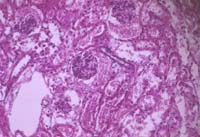
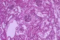
Instructions for Koehler illumination| STEP 1 Focus your sample in brightfield. (Note the dark shadow in the upper right) |
 |
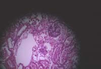
| STEP 2 Close the field diaphragm so it looks something like this: |
 |
| STEP 3 Focus the edge of the diaphragm by adjusting the condenser height, so it looks like this: (if the image moves out of your field of view, skip to step 4, then come back to step 3) |
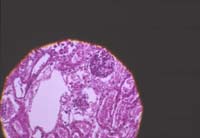 |
| STEP 4 center the image using the two centering screws, so it looks like this: (Note centered, crisp edge) |
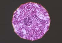 |
| STEP 5 Open the field diaphragm until it is at the edge of the field of view. (Note that the shadow in step 1 is gone.) |
 |
Contrast can be adjusted using the CONDENSER diaphragm.
However, be careful when adjusting the condenser diaphragm. Closing the condenser diaphram reduces resolution. To maximize both contrast and resolution, close the diaphragm just to the point where the image begins to get dark and no further. This position is especially important when using Nomarski optics.
![]()
![]()
last revised 27 Nov 2000 by mc; previous authoring by cbm