
| |
These are the first fluorescent images we took with the new fluorescent attachment to the STEMI.
From John Greally's lab:
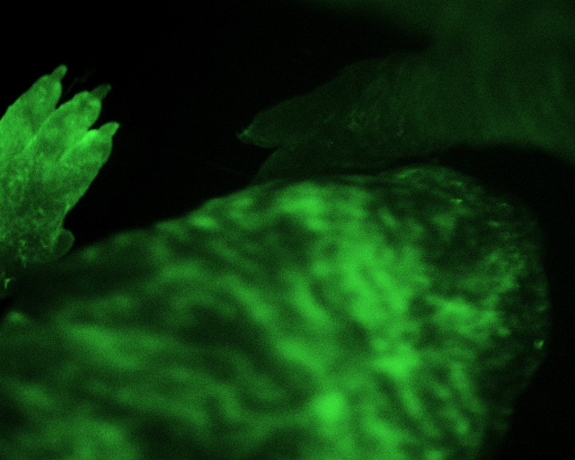
D4/XEGFP mice crossed with CSTBL/6 (originally created by Dr. Andras Nagy).
These female mice (5 days old) have an X linked GFP transgene that undergoes maturation in
half the cells resulting in a variegated fluorescence.
The pictures were taken with a 1.6X objective. Also the microscope has a 1X and a
0.6X objective so that it should be able to image a larger field than shown here.
Here is the top of a mouse's head:
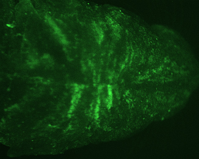
The mouse on the bottom is GFP expressing. The mouse in the top right corner is
barely visible because it is GFP negative.
The paw on the left is of a GFP negative mouse. The paw on the right is of a GFP
expressing mouse.
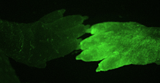
After the mice were imaged they were returned to their mother for further maturation and breeding.