
| |
This is pollen from a tree in the flowerboxes outside Low Housing on campus here.
Using the Zeiss dissection microscope, we could take macro images (two color images at left and more here.).
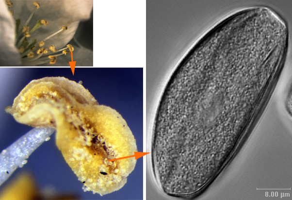
Using the Leica AOBS microscope, we could scan at high resolution.
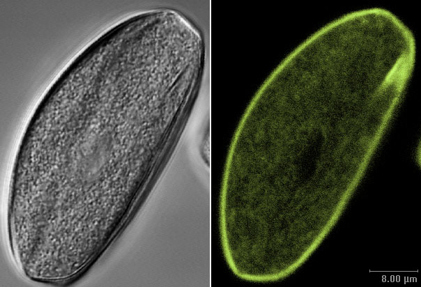
The pollen may be imaged by its own fluorescent emission.
When this pollen is excited with 405 nm light, this is the resulting spectrum:

However, when exciting at 365 nm +/- 20 nm, we could see by eye that each grain had a
distinct red line running through it.
This was not detected by the confocal with excitation by a single line at 40 nm longer.
Here is a series of thin optical sections taken through another grain.
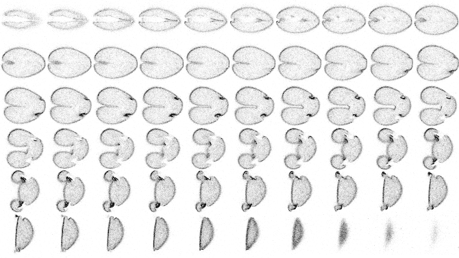
Click here for a 2.46MB movie of the montage above.
Although the corners of the grain cracked (probably due to rough handling), it shows the
basic 3D structure.
All material on this page © 2004 all right reserved by Michael Cammer and the Albert Einstein College of Medicine of Yeshiva University