
| |
| Leica AOBS confocal microscopy |
A few early (at installation in 2002) example images.
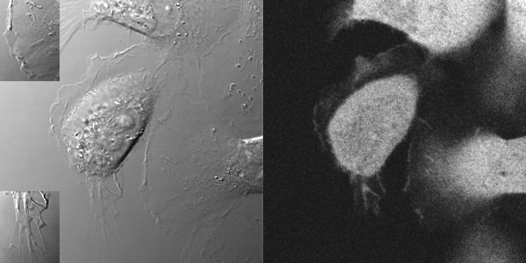
Multiple fluorescent probes can be collected with a DIC image to see overall cell
structure.
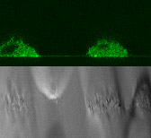
XZT imaging is a standard function. This is a side view of live GFP expressing
cells.
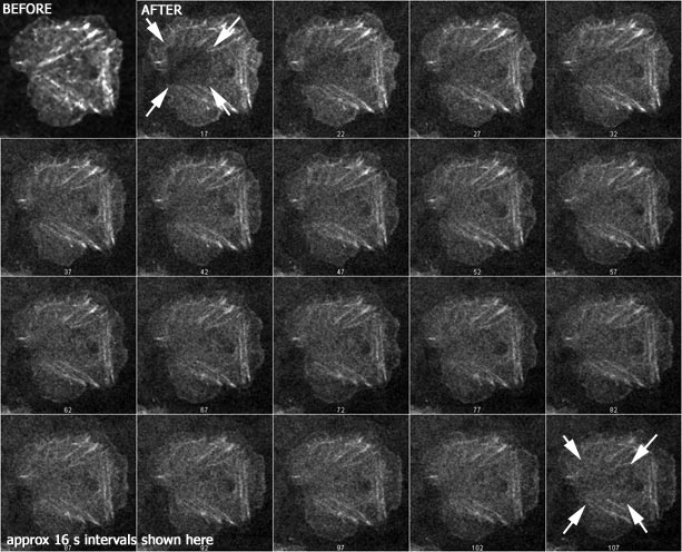
FRAP is standard on this instrument.
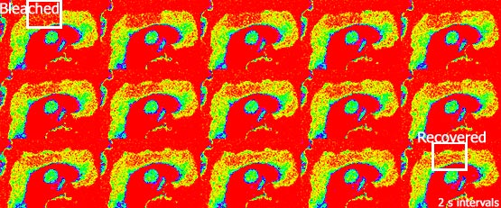 and
and
Sample method section for a paper:
Images were collected with a Leica TCS SP2 AOBS confocal microscope (Mannheim, Germany) with 25X and 60X oil immersion optics. Laser lines at 488nm and 543nm for excitation of Cy2 and Cy3 were provided by an Ar laser and a HeNe laser. Detection ranges were set to eliminate crosstalk between fluorophores.
For more information on confocal at AECOM, please contact Michael at cammer@aecom.yu.edu.