
| |
| Example of Manual Area Measurement: |
The point of this web page is to show that cells in culture (or in vivo) can be imaged at discrete sequential times and measurements about speed, shape, size or other morphometrics can be made.
Cells were imaged with a 20X phase contrast objective on an Olympus IX 70 inverted microscope with an environmental chamber. A custom script was written for I.P. Lab Spectrum to control a shutter and camera on the microscope. Images were collected every minute and saved to disk.
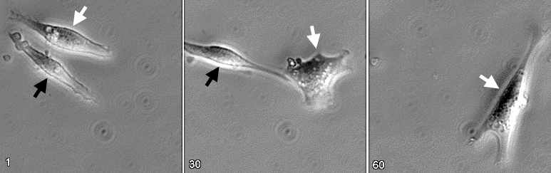 Phase contrast images of cells at first timepoint, middle timepoint, and final timepoint. The white arrows point to the cell that was traced and measured (see below). |
Using a set of custom macros written at the AIF for use with NIH-Image, the user traced the cell at each timepoint. One of the macros drew the following perimeter trace:
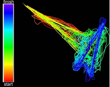
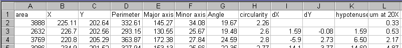
The macros include measurement commands so that data could be loaded into a spreadsheet for subsequent analysis.
Locations of the cell center (or "centroid") were plotted.
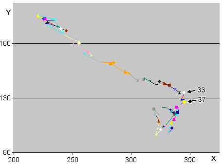
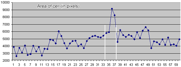
Timepoints 33 and 37 are marked in the centroid paths above. At timepoint 33 the cell ceases a persistent walk at about 30 degrees towards the lower right of the image. The cell changes direction. Note in the plot of cell area below that the cell area increases dramatically between timepoints 33 and 37 (marked by white vertical lines). This spreading is associated with the change of direction and could be quantitated for a population of cells as well as for our lone anecdotal cell.
In summary, direction, speed, and area can be easily quantified. This page does not show quantitation of persistence or measurement of angular change, but these can be calculated from the data. Also, measurements for relative distances for features within the cells can be made. In addition, were the cells fluorescent, measurements could be made with respect to the intensities overall or in specific compartments and could be correlated with the morphometric data. And, perhaps, the cells could be measured automatically instead of by manual tracing.
![]()
last revised 21 Feb 2001 by mc