| |
| Basic IRM. |
Example of internal reflectance microscopy with the BioRad Radiance 2000 confocal.
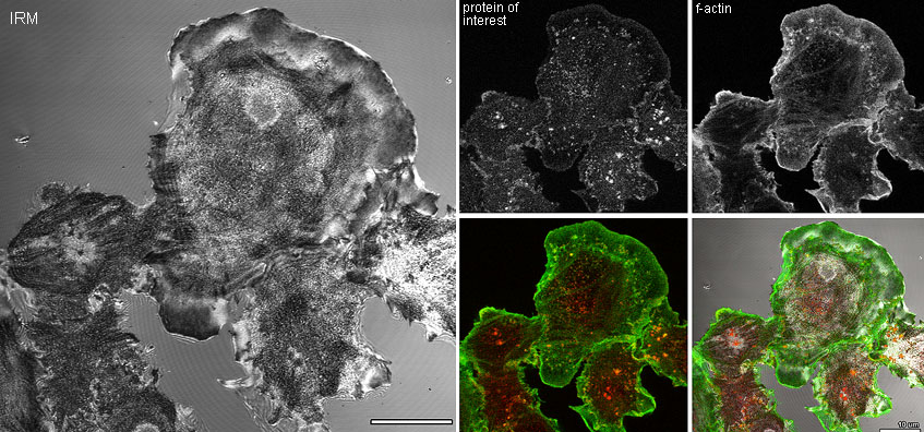
| In the example below, cells were plated on coverslips,
fixed, and stained for f-actin and another protein of interest. The IRM image shows the cell adherent to or pressing against the substrate as black. Note that many of the red dots, especially towards the center of the cell, appear to be associated with contact points. Data, such as these, can be quantitated. Simultaneous IRM imaging with two fluorescent probes and a transmitted light image (Nomarski or phase contrast -- not show here) is standard with the BioRad Radiance 2000 confocal.
|
 |
| IRM imaging of live cells is especially useful for studying the dynamics of cell adhesion or more general motility or locomotion. See Bailly, M., Yan, L., Whitesides, G.M., Condeelis J.S. and Segall, J.E. (1998) Regulation of protrusion shape and adhesion to the substratum during chemotactic responses of mammalian carcinoma cells. Experimental Cell Research 241(2):285-299. [Related movies.] [Alternate version of movie.] |
More examples of IRM and IRM on the confocal vs. IRM with a 50/50 mirror on a conventional epi-illumination microscope here.
How to set the optics for IRM or any reflection microscopy on the BioRad Radiance 2000.
The idea is to zap the sample with a laser line and only allow that laser line to go back to the PMT. Tricks include keeping the iris diaphragm closed more than the ideal calculated by the software.
Reflection imaging does involve some scattering and maybe a hot spot in the center of the image. Both are not desireable. BioRad can provide a polarizing (or is it a 1/4 wave plate??) filter to be inserted in the light path which eliminates these contrast masking problems.
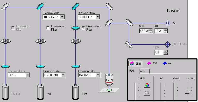
In its simplest form, zap with 488 nm light and
collect only the 488 nm light.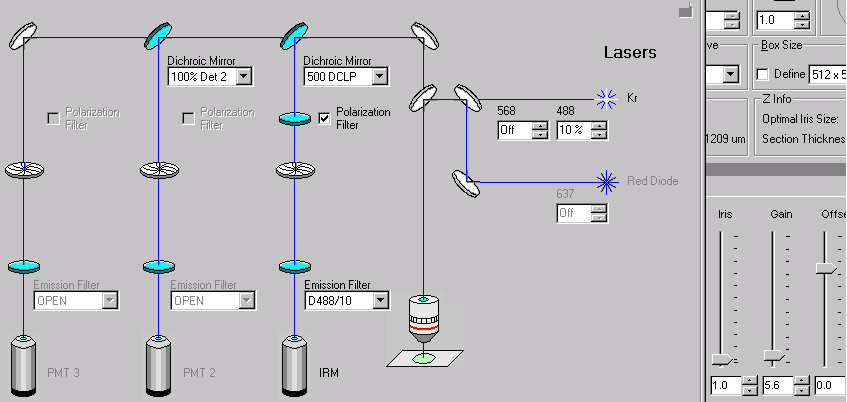 The Dichroic mirror
could be set for anything except "100% Det 2/3". Even if you don't have the correct bandpass filters in front of the
other PMTs, the dichroic can be used alone for some IRM and fluorescent simultaneous
imaging. Similary, IRM can be done in other PMTs than #1. Here's a simple
example of IRM and red fluorescence imaging simulataneously:
|
Comments, questions or suggestions? Contact Michael Cammer