
| |
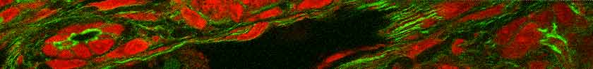
The confocal microscope is for 1.) thin optical sections of fluorescence stained material, 2.) thin optical sections of reflectance stained material or 3.) reflectance imaging of relief surfaces or thin spaces such as the cell-substrate interface.
The confocal microscope is not for convenient multiple color imaging; we have widefield microscopes for this. The confocal microscope is not for low light imaging;
we have widefield microscopes for this. Probes for BioRad Radiance 2000 Confocal Microscopy:| Main Manual Checklist for Startup and Shutdown |
| Recommendations for slide preparation for the confocal. |
Information for methods sections:
Nikon Eclipse 200 modified laser safe (Melville, NY). Infinity corrected 10X, 20X, 40X, 60X objectives.
BioRad Radiance 2000 laser scanning confocal microscope (CA).
Kr/Ar laser for excitation at 488 and 568 nm and diode laser for excitation at 633 nm.
Standard filters with narrow emissions for imaging green and red channels. Far red
imaged with 650LP filter.
Absence of bleed through from one wavelength to a longer wavelength
is achieved using one of these methods:
1. balancing of laser intensities with gain settings;
2. "lambda scanning" which is scanning each line rapidly in sequence with
individual excitations; or
3. sequential imaging of each dye by excitation with a single laser line.
 |