 |
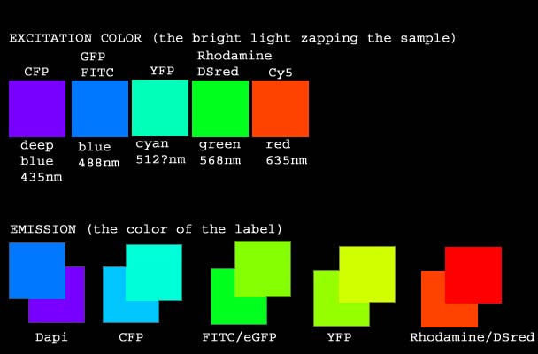
For instance, let's say your have cultured cells or a vibratome
section of tissue and you want to look at DNA, f-actin, your favorite protein and
microtubules (tubulin) by fluorescent light microscopy. We might suggest you use
respectively DAPI (binds to DNA), fluorescein
(bound to phalloidin which binds to f-actin filaments), Cy3
(antibody method for your favorite protein) and Cy5 (antibody
method to alpha or beta tubulin). In general, you
may use any one dye from each of the groupings below for multi-labeling -- this list is
not exhaustive (l is the emission color):
| COLOR OF DYE |
WE RECOMMEND |
ALSO OK |
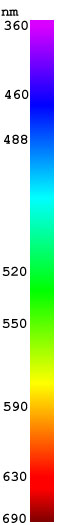
| Blue, approximately 460nm. |
Dapi, Hoechst |
Cascade Blue, Pacific Blue, FluoroBlue |
| Green, approximately 520nm |
Alexa 488, Cy2,GFP, eGFP, YFP, Fluo3 |
FITC (fluorescein), Oregon Green, , Bodipy, NBD,
Calcium Green |
| Red, approximately 590nm. |
Alexa 568, Cy3, Propidium iodide, Mitotracker red,
DiI, DiA |
Rhodamine, Texas red, TRITC, Alexa 594, Alexa 546,
DsRed2 |
| Near infra red, approximately 670nm |
Cy5, Alexa 633, Alexa 647 |
DRAQ5, APC |
The following table shows the filters we have
for Olympus microscopes:
| NAME OF FILTER |
Excitation |
Dichroic |
Emission |
approximate color |
| DHE (Dehydroergosterol) |
D335/20 |
365dclp |
D405/40 |
|
| Dapi |
360 |
|
400 LP |
|
| CFP |
436 |
455 |
480 |
|
| FM143 |
480/30 |
570 LP |
505dcxt |
|
| FITC |
460-590 (depends on exact set) |
500 |
510+ BP or LP |
|
| YFP |
500 |
515 |
535+ BP or LP |
|
| Rhodamine |
550-580 (depends on exact block) |
|
|
|
| Cy5 |
620/60 |
660LP |
700/75 |
|
| Custom filters can be purchased and there may be
more filters available since this web page was last updated. |
|
For questions about GFP, mCherry, etc.
Please visit the Fluorescent
Protein Resource Center.
N.b. FITC, GFP and other green
labels cannot be imaged with DsRed!
Probes for BioRad Radiance 2000 Confocal
Microscopy:
For the BioRad confocal microscope, you
may not use any of the stains from group blue
which require excitation at 360 nm. You may use any of the dyes from the green, red, or
near infra red categories. The confocal
microscope has lasers that excite dyes at 488 nm, 568 nm and 637 nm only. [Example triple label.]
|
Probes for Leica AOBS Confocal Microscopy:
The Leica AOBS uses variable spectral detection instead of
traditional emission filters. There are a few laser lines for excitation and the
dyes are limited by these. Any dye that is excited at one of these following
wavelengths can be used.
405; 458; 476; 488; 514; 543; and 633 nm.
N.b. that there is no line at 568 which is the best line for reds similar to rhodamine. |
An often asked question is, What is good
for labeling nucleic acids other than DAPI?
This is an important question because Dapi & Hoechst cannot be used with the
confocal or with CFP.
The first answer we provide is propidium iodide in the red range because
it is so easy to use.
But there is a new family of nucleic acid stains by Molecular Probes called Syto.
Nobody has used them in the AIF so we can't recommend them, but we'd love somebody to try
and show us the results. The most promising Syto dyes appear to be Syto 13 or Syto 16 in the green range or Syto
60 in the infra red range. |
Suggestions for work with live cells:
When FITC is excited, it produces free radicals which can be very harmful to live cells.
Also, FITC is pH sensitive such that its intensity changes depending on the pH.
And FITC bleaches very quickly. For these reasons, we recommend more stable
and brighter Alexa 488 or Cy2 instead of FITC. Or, if you can, switch to a red probe
such as Alexa 568, Cy3, Rhodamine, or Texas Red. |
On the digital stations, any custom filters can be
built for any wavelenths between 360 nm and 700+ nm.
For instance, we have special filters on Digital Station #1 for
double labeling and time lapse imaging of cyanFP
and yellowFP. Digital Station #3 has a few
special filters, for instance for chromosome painting. And the Leica AOBS confocal can image any part of the visible
spectrum without using filters. |

 Comments:
 More Resources: